Corneal Dystrophy in a Pomsky
It's incredibly worrying when you notice something isn't quite right with your Pomsky's eyes. That sudden cloudiness, a subtle change in texture, or even just a general "off" look can send any pet parent into a spiral of concern, wondering if their beloved companion is in pain or losing their vision. This comprehensive guide will walk you through everything you need to know about corneal dystrophy in Pomskies, from understanding its nature to diagnosing and managing it, ensuring you’re equipped to support your furry friend.
Understanding Corneal Dystrophy: What It Is Exactly?
The term "corneal dystrophy" can sound quite intimidating, but at its heart, it refers to a group of inherited, non-inflammatory conditions that affect the cornea, the clear, outermost layer of your dog's eye. Unlike injuries or infections, these conditions typically aren't caused by external factors but rather by genetic predispositions. For Pomsky owners, understanding this distinction is crucial because it informs how the condition might progress and what management strategies are most effective.
The Cornea and Its Vital Role
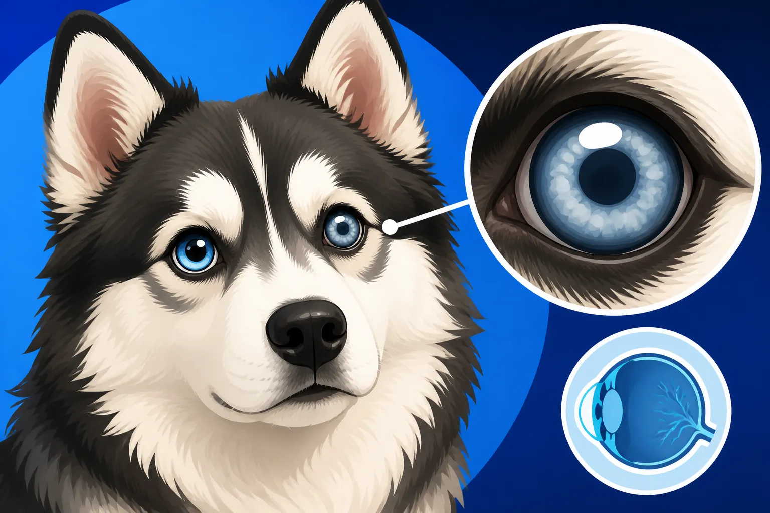
Imagine looking through a dirty or scratched window; everything becomes blurry and distorted. Now, imagine that window is a fundamental part of your vision. That’s essentially what the cornea is for your Pomsky: the clear, dome-shaped front surface of the eye that acts as its primary lens. Its job is to protect the inner structures of the eye and to refract (bend) light, focusing it onto the retina at the back of the eye. For clear vision, the cornea must be perfectly transparent.
The cornea itself is made up of several distinct layers: the outer epithelial layer, the middle stromal layer, and the inner endothelial layer. Each layer plays a specific role, and when any one of them develops a dystrophy, its transparency can be compromised. This can lead to cloudiness, changes in light transmission, and, in some cases, discomfort or impaired vision. A healthy, sparkling eye is a sign of good general well-being, and any changes to that clarity are definitely worth investigating.
Types of Corneal Dystrophy in Dogs
Corneal dystrophy isn't a single condition but rather an umbrella term for several distinct types, each affecting a different layer of the cornea. Knowing the type can help your vet determine the best course of action and what to expect in terms of progression.
- Epithelial Corneal Dystrophy: This type affects the outermost layer of the cornea. It often manifests as small, grayish-white, often circular or oval opacities that can appear anywhere on the cornea. Sometimes, these can be quite subtle and might only be visible upon close inspection or with specialized equipment. In more severe cases, recurrent erosions or ulcers can develop, which are not only painful but also make the eye vulnerable to infection.
- Stromal Corneal Dystrophy: This is probably the most commonly recognized type and affects the middle, thickest layer of the cornea. It's often characterized by bilateral (affecting both eyes), non-inflammatory, crystalline or milky opacities. These opacities are deposits of cholesterol or other lipids within the corneal stroma. They can vary significantly in size and density, from faint hazes to dense, sparkling white spots that can sometimes look like tiny shards of glass embedded in the eye. While often striking in appearance, many dogs with stromal dystrophy retain good vision, especially if the deposits are not directly over the pupil.
- Endothelial Corneal Dystrophy: This type affects the innermost layer of the cornea, responsible for pumping fluid out of the cornea to keep it clear and prevent swelling. When this layer malfunctions, fluid accumulates within the cornea, leading to a blue-gray haze known as corneal edema. This edema often starts at the periphery of the cornea and slowly progresses inwards, eventually covering the entire surface. While initially it might not cause pain, chronic edema can lead to painful corneal blisters (bullae) that can rupture, causing ulcers.
For Pomskies, given their parentage, particular attention is often paid to the stromal type due to breed predispositions in some Huskies, though any form can potentially arise.
Differentiating From Other Eye Conditions
It's easy to jump to conclusions when you see cloudiness in your Pomsky's eye. Is it a cataract? Glaucoma? An injury? It's important to remember that corneal dystrophy is distinct from these other common eye ailments, although some symptoms might overlap, making a veterinary diagnosis essential.
Cataracts, for instance, involve clouding of the lens inside the eye, not the cornea itself. While both can cause a hazy appearance, a vet can easily differentiate between them with an ophthalmoscope. Glaucoma, on the other hand, is characterized by increased pressure inside the eye, which can lead to corneal edema (a blue haze) and pain. However, glaucoma is often very painful and progresses rapidly, whereas corneal dystrophy is typically non-painful (unless complicated by ulcers) and progresses slowly, if at all. Injuries, infections, or inflammatory conditions like uveitis also cause cloudiness, redness, and usually significant discomfort or discharge. The key difference with corneal dystrophy is its non-inflammatory, often bilateral, and symmetrical nature, combined with its hereditary basis. Your vet will perform specific tests to rule out these other possibilities, ensuring your Pomsky gets the correct diagnosis and management plan.
Recognizing the Signs: How Corneal Dystrophy Shows Up in a Pomsky
Observing your Pomsky closely is the first step in noticing any changes, especially with their eyes. The subtle signs of corneal dystrophy can be easy to miss in the early stages, often requiring a keen eye and regular interactions with your furry friend. Since Pomskies are known for their striking eyes, any alteration will likely catch your attention.
Early Symptoms and Subtle Changes
The most classic sign of corneal dystrophy in a Pomsky is a noticeable cloudiness or opacity in one or both eyes. This isn't usually a sudden onset like an injury but a gradual development. You might notice:
- White or Grayish Patches: These can appear as small, often circular or oval, lesions on the surface or deeper layers of the cornea. In some stromal cases, they can even look like sparkling crystals. They might be faint initially, becoming more prominent over time.
- A "Milky" or Hazy Appearance: The clarity of the cornea can diminish, giving the eye a slightly dull or milky look, rather than the bright, reflective surface you're used to seeing.
- Changes in Eye Texture: The surface of the eye might appear uneven or rough in cases of epithelial dystrophy, especially if recurrent erosions are forming.
- No Obvious Pain or Redness (Initially): Unlike an infection or injury, corneal dystrophy usually doesn't cause a red, watery, or squinting eye in its early stages. This lack of acute distress can sometimes lead owners to delay a vet visit, mistaking the cloudiness for aging or a minor irritation.
It's important to remember that many forms of corneal dystrophy are not painful, which can be both a blessing and a curse. While your dog isn't suffering, the lack of overt pain signals might mean the condition progresses unnoticed for a while. Regular eye checks at home, looking at your Pomsky's eyes in good light, can help you catch these subtle changes early.
Breed-Specific Predispositions
Understanding the parent breeds of your Pomsky is key when considering any genetic health condition. Pomskies inherit traits from both Siberian Huskies and Pomeranians, and unfortunately, this can include genetic predispositions to certain eye conditions.
Siberian Huskies, for example, are known to have a higher incidence of several ocular issues, including certain types of corneal dystrophy, such as crystalline corneal dystrophy (a form of stromal dystrophy). They can also be prone to conditions like The Juvenile Cataracts a Pomsky Gets From the Husky or progressive retinal atrophy. Pomeranians, while generally having fewer widespread hereditary eye conditions, can still contribute to the overall genetic makeup that influences a Pomsky's susceptibility. This mixed heritage means that your Pomsky could potentially inherit predispositions from either side. This is why when you're looking at a Pomsky's overall health profile, you have to consider The Two Opposite Disease Lists a Pomsky Inherits. Responsible breeders will screen their breeding dogs for known genetic issues to minimize the risk of passing them on, but it's not always a guarantee given the complexity of genetics.
When to Seek Veterinary Attention
Any change in your Pomsky's eyes warrants a trip to the vet, but some situations are more urgent than others. While corneal dystrophy typically isn't an emergency, complications can arise, and other conditions mimic its appearance.
Urgent Veterinary Attention (within 24 hours):
- Your Pomsky is squinting, rubbing their eye, or showing obvious signs of pain.
- The eye appears suddenly red, swollen, or has thick discharge.
- Vision seems acutely impaired (e.g., bumping into things, hesitant to move).
- The cloudiness or haze develops very rapidly.
Routine Veterinary Visit (within a few days to a week):
- You notice a new, subtle cloudiness, white spot, or change in clarity that isn't accompanied by pain or redness.
- You observe recurrent watery eyes or mild irritation without other severe symptoms.
- You just want a professional opinion on anything that seems "off" with their eyes.
It’s always better to err on the side of caution. Even if it turns out to be nothing serious, a vet check-up provides peace of mind. Plus, early detection of any eye condition, including corneal dystrophy, can lead to more effective management and prevent potential complications down the line. Remember, your Pomsky can't tell you if their eye is bothering them, so you are their first line of defense!
The Diagnostic Journey: Pinpointing Corneal Dystrophy
When you suspect an eye issue with your Pomsky, your local veterinarian is usually the first point of contact. They can perform an initial assessment and, if necessary, refer you to a veterinary ophthalmologist for specialized diagnostics. The diagnostic process aims to accurately identify the condition, rule out other possibilities, and establish a baseline for monitoring.
Your Vet Visit: Initial Examination and Basic Tests
When you bring your Pomsky in for an eye concern, your general practitioner will likely start with a thorough physical examination, including a careful inspection of the eyes. This initial exam is crucial for distinguishing simple irritations from more serious underlying conditions.
Typical tests performed during a routine vet visit for eye issues include:
- Ophthalmoscopic Examination: Your vet will use an ophthalmoscope, a specialized light and magnifying tool, to look directly into your Pomsky's eye, examining the cornea, lens, retina, and optic nerve. This helps them determine the exact location of any opacity and assess the overall health of the eye.
- Schirmer Tear Test: This test measures tear production to rule out "dry eye" (keratoconjunctivitis sicca), which can cause corneal irritation and cloudiness. A small strip of filter paper is placed inside the lower eyelid for one minute.
- Fluorescein Stain Test: A special orange dye (fluorescein) is applied to the eye. This dye adheres to any areas where the outermost corneal layer (epithelium) is damaged, appearing bright green under a blue light. This helps the vet identify corneal ulcers or abrasions, which are not typically part of primary corneal dystrophy but can be a complication.
- Pupillary Light Reflex (PLR): Testing how your Pomsky's pupils react to light gives insight into neurological function and retinal health.
These initial tests provide your vet with a great deal of information and can often differentiate corneal dystrophy from more acute problems like infections or injuries.
Specialized Ophthalmic Diagnostics
If your general vet suspects corneal dystrophy, or if the case is complex, they might recommend a referral to a board-certified veterinary ophthalmologist. These specialists have advanced equipment and expertise to provide a more precise diagnosis.
Specialized ophthalmic diagnostics might include:
- Slit Lamp Biomicroscopy: This advanced microscope allows the ophthalmologist to examine the cornea in high magnification and detail, layer by layer. It can precisely identify the type and depth of corneal opacities, which is critical for differentiating between epithelial, stromal, and endothelial dystrophies.
- Tonometry: This measures the intraocular pressure (IOP) to check for glaucoma. Glaucoma can cause corneal edema, which might look similar to endothelial dystrophy, but the underlying cause and treatment are very different.
- Corneal Scraping or Biopsy: In rare cases, if there's an unusual appearance or concern about other conditions, a small sample of corneal cells might be taken for microscopic examination (cytology) or a biopsy for histopathology. This can rule out other infiltrative diseases or certain types of infections.
- Electroretinography (ERG): If there's concern about retinal function or vision loss, an ERG can assess the electrical activity of the retina. This is more common when looking at inherited retinal diseases rather than primary corneal dystrophy, but it may be part of a comprehensive eye exam, especially if there are other concerns about The Eyes a Pomsky Gets From the Husky.
The ophthalmologist’s assessment will provide a definitive diagnosis, give you a clearer picture of the prognosis, and help formulate the most appropriate long-term management plan for your Pomsky.
Ruling Out Other Eye Concerns
One of the most critical aspects of diagnosing corneal dystrophy is ruling out other conditions that can present with similar symptoms, particularly corneal cloudiness. A thorough diagnostic process ensures your Pomsky receives appropriate care and prevents unnecessary or incorrect treatments.
Conditions that often need to be differentiated from corneal dystrophy include:
- Corneal Edema from Glaucoma: As mentioned, elevated eye pressure in glaucoma can cause the cornea to swell and appear hazy. However, glaucoma is often painful and rapidly progressive, requiring immediate intervention to save vision.
- Corneal Ulcers or Injuries: Traumatic injuries, scratches, or foreign bodies can cause localized cloudiness, redness, and significant pain. The fluorescein stain test is highly effective in identifying these.
- Keratitis (Corneal Inflammation): This can be caused by infections (bacterial, viral, fungal), allergies, or irritants. Keratitis usually involves inflammation, redness, pain, and discharge, which are not typical of uncomplicated corneal dystrophy.
- Pigmentary Keratitis: This involves melanin pigment migrating onto the cornea, often due to chronic irritation or dry eye. It appears as dark brown or black spots, distinct from the white/grey opacities of dystrophy.
- Cataracts: This is clouding of the eye's lens, not the cornea. While both can cause vision impairment, their location within the eye is different and easily distinguished by a vet. For example, The Juvenile Cataracts a Pomsky Gets From the Husky is a distinct condition that affects the lens.
- Uveodermatologic Syndrome: This rare autoimmune condition can affect the eyes, skin, and coat, sometimes causing ocular inflammation and secondary corneal changes. It's a much broader systemic disease that also affects pigmentation, and understanding it is part of why you'd want to consider Whether a Pomsky Gets Uveodermatologic Syndrome in a differential diagnosis.
The meticulous process of elimination performed by your vet and potentially a veterinary ophthalmologist ensures that any cloudiness or changes in your Pomsky's eyes are correctly identified, allowing for the most targeted and effective care.
Living with Corneal Dystrophy: Management and Treatment Options
Once your Pomsky has been diagnosed with corneal dystrophy, the focus shifts to management. Since corneal dystrophy is often a genetic condition, there isn't typically a "cure," but rather strategies to minimize symptoms, prevent complications, and maintain your dog's quality of life. The approach will vary depending on the type and severity of the dystrophy.
Medical Management: Eye Drops and Topical Treatments
For many forms of corneal dystrophy, medical management, primarily through topical eye drops, is the cornerstone of treatment. The goals are often to keep the eye comfortable, hydrated, and to address any secondary issues.
- Lubricating Eye Drops or Gels: These are perhaps the most common recommendation, especially for epithelial dystrophies or any type where the corneal surface might be compromised. Regular application helps keep the eye moist, reduce irritation, and support the integrity of the corneal surface. Brands like Refresh Optive Mega-3 or Genteal Gel are frequently recommended by vets.
- Hypertonic Saline Ointment or Drops: For endothelial corneal dystrophy, where fluid accumulates in the cornea causing edema, hypertonic (5% sodium chloride) saline can be used. This draws excess fluid out of the cornea, helping to reduce swelling and improve clarity. It doesn't cure the underlying problem but manages the symptoms.
- Topical Anti-Inflammatory Medications: If there's any associated inflammation or irritation, or if secondary conjunctivitis develops, your vet might prescribe mild topical steroids or non-steroidal anti-inflammatory drugs (NSAIDs) for a short period. This must be done cautiously and under strict veterinary guidance, as steroids can worsen existing ulcers.
- Antibiotic Eye Drops: If recurrent corneal erosions or ulcers develop (which can happen with epithelial dystrophy or severe endothelial dystrophy), antibiotic drops or ointments will be prescribed to prevent or treat bacterial infections.
Consistency is absolutely key with eye medications. Missing doses can set back progress or lead to complications. It can be challenging to administer drops to a wiggly Pomsky, but with patience, positive reinforcement, and tasty treats, most dogs can learn to tolerate it.
Surgical Interventions and Advanced Therapies
While medical management is usually the first line of defense, some cases of corneal dystrophy, particularly those causing significant pain, recurrent ulcers, or severe vision impairment, might warrant surgical intervention.
- Superficial Keratectomy: This procedure involves surgically removing the affected superficial layers of the cornea. It's sometimes considered for epithelial or superficial stromal dystrophies that cause severe, recurring erosions or painful, visually significant opacities. The goal is to remove the diseased tissue and allow healthy corneal tissue to regenerate. The success rate varies, and recurrence is possible.
- Grid Keratotomy or Diamond Burr Debridement: These techniques are used to treat recurrent corneal erosions that often accompany certain epithelial dystrophies. They involve making tiny scratches or using a fine burr on the corneal surface to promote better adhesion of the epithelial cells, helping the cornea heal more effectively and prevent future erosions.
- Conjunctival Flap Surgery: In cases of chronic, non-healing ulcers resulting from corneal dystrophy complications, a flap of conjunctiva (the pink tissue lining the eyelids) can be surgically attached over the ulcer. This provides blood supply and support to the healing cornea.
- Corneal Transplant: While rare and technically challenging in veterinary medicine, corneal transplantation might be considered in very severe, vision-threatening cases of stromal dystrophy. This involves replacing the diseased cornea with healthy donor tissue.
These surgical options are typically performed by a veterinary ophthalmologist and involve significant costs and recovery periods. They are usually reserved for situations where medical management is no longer sufficient to maintain the dog's comfort and quality of life.
Home Care and Environmental Adjustments
Beyond medications and potential surgeries, simple home care and environmental adjustments can play a significant role in managing corneal dystrophy and supporting your Pomsky's eye health.
- Gentle Eye Cleaning: If there's any discharge or crusting, gently wipe around your Pomsky's eyes with a soft, damp cloth or a veterinary-approved eye cleaning solution. Always wipe away from the eye, and use a separate clean cloth for each eye to prevent cross-contamination.
- Reduce Irritants: Minimize exposure to environmental irritants like dust, smoke, strong cleaning chemicals, or excessive wind. If your Pomsky enjoys car rides with their head out the window, consider investing in Doggles or protective eyewear.
- Avoid Rubbing: If your Pomsky is rubbing or scratching their eyes, this can worsen the condition or cause secondary ulcers. An Elizabethan collar (e-collar) might be necessary, especially if they are prone to scratching.
- Comfortable Environment: Ensure your Pomsky has a comfortable, calm place to rest. If vision is somewhat impaired, keep their living environment consistent to help them navigate easily. Don't rearrange furniture frequently.
- Regular Observation: Continue to closely monitor your Pomsky's eyes daily. Look for any new cloudiness, redness, discharge, or signs of discomfort. Jot down any changes to discuss with your vet.
These practical steps, combined with veterinary treatments, form a comprehensive approach to managing corneal dystrophy and ensuring your Pomsky remains as comfortable and happy as possible.
Prognosis and Long-Term Impact on Your Pomsky
Receiving a diagnosis of corneal dystrophy can be unsettling, but it's important to understand the typical prognosis and what the long-term impact on your Pomsky's life might be. Most forms of corneal dystrophy are not life-threatening, but they do require ongoing management.
Vision Impairment and Quality of Life
One of the primary concerns for any eye condition is its effect on vision. For many Pomskies with corneal dystrophy, especially the stromal types, the impact on vision is often minimal. The opacities might be noticeable to you, but they may not significantly impede your dog's ability to see, particularly if the deposits are not directly over the pupil. Most dogs adapt incredibly well to minor vision changes, relying more on their excellent sense of smell and hearing.
However, certain types of corneal dystrophy, particularly severe endothelial dystrophy leading to chronic edema and bullae, or extensive stromal dystrophy, can lead to more significant vision impairment. If recurrent corneal erosions are a problem, they can cause pain and temporary vision blurriness. It's rare for corneal dystrophy alone to cause complete blindness, but it is a possibility with very severe, unmanaged cases, or if complications like deep ulcers or secondary infections arise. Thankfully, Whether Corneal Dystrophy Blinds a Pomsky is usually not the case. The overall quality of life for a Pomsky with corneal dystrophy is generally good, provided the condition is monitored and managed appropriately. They can continue to enjoy walks, play, and cuddle just like any other dog.
Monitoring and Regular Check-ups
Consistent monitoring and regular veterinary check-ups are paramount for any Pomsky diagnosed with corneal dystrophy. The frequency of these visits will depend on the type and stability of the condition, but generally, yearly eye exams are a good idea. This is part of The Screening a Pomsky Should Have Yearly every Pomsky should undergo.
During these follow-up appointments, your vet or veterinary ophthalmologist will re-examine the eyes, assessing the progression of the dystrophy, checking for any new opacities, and ensuring no secondary complications (like ulcers or infections) have developed. They might repeat some diagnostic tests, such as slit lamp examination or tonometry, to compare findings to previous visits. It's also an opportunity for you to discuss any changes you've observed at home, such as increased rubbing, redness, or perceived changes in vision.
Beyond professional exams, you are your Pomsky's best advocate. Make it a habit to check their eyes daily. Look for any new cloudiness, redness, discharge, or signs of discomfort. Notice if they're bumping into things more often or if their behavior suggests they're struggling with vision. Early detection of any worsening or new problem can make a huge difference in the outcome. Just like wondering Whether a Pomsky Needs a Yearly Eye Exam for general health, regular eye-specific checks are crucial. It's also important to remember that this condition is separate from others that might cause vision loss, like Whether a Pomsky Goes Blind From Cataracts, which affects the lens rather than the cornea.
Financial Considerations and Pet Insurance
Managing a chronic condition like corneal dystrophy, especially if it requires specialist visits, ongoing medications, or potential surgery, can unfortunately lead to significant veterinary expenses. Diagnostic tests, specialist consultations, and long-term medications can add up quickly.
Consider the potential costs:
- Initial Veterinary Visit & Diagnostics: $100 – $300
- Specialist Ophthalmology Consultation: $150 – $400 (initial exam)
- Specialized Tests (Slit Lamp, Tonometry, etc.): $100 – $300+ per session
- Ongoing Medications (Drops/Ointments): $20 – $80+ per month, depending on the medication
- Surgical Procedures (if needed): $1,000 – $5,000+ per eye, depending on the complexity
These costs highlight the value of pet insurance. Having a robust pet insurance plan, like one from Healthy Paws, can be an absolute lifesaver when those specialist bills start rolling in. Policies from providers like Healthy Paws can help cover a significant portion of diagnostic tests, specialist fees, medications, and even surgery for unexpected illnesses or accidents. It’s always wise to explore options like Healthy Paws pet insurance to help mitigate these unexpected costs, allowing you to focus on your Pomsky's health without financial stress being the primary concern. Always review policy details carefully to understand what is covered, as pre-existing conditions are typically excluded.
| Symptom/Sign | What it Might Look Like | Potential Meaning (in Corneal Dystrophy) | Action to Take |
|---|---|---|---|
| White/Grayish Haze | Faint film, cloudy spots, or crystalline sparkle | Deposits in cornea (stromal or epithelial dystrophy) | Consult vet; often not urgent if no pain |
| Blue-Gray Cloudiness | General blue tinge, especially at edges | Corneal edema (endothelial dystrophy or glaucoma) | Urgent vet visit if rapid onset or pain |
| Squinting/Rubbing | Eye closed partially, pawing at face | Pain, irritation (recurrent erosion, ulcer, foreign body) | Urgent vet visit |
| Redness/Discharge | Bloodshot eyes, watery or thick discharge | Inflammation, infection (secondary complication) | Urgent vet visit |
| Bumping into Objects | Disorientation, hesitation to jump | Significant vision impairment or loss | Urgent vet visit to assess vision loss |
| No Visible Changes | Eyes appear clear, no discomfort | Healthy eyes or very early, subtle dystrophy | Regular home checks, yearly vet eye exam |
Breeding Practices and Genetic Responsibility
Corneal dystrophy, by definition, is a hereditary condition. This means it's passed down from parent dogs to their offspring. For Pomskies, a relatively new designer breed, understanding the genetic aspect and the importance of responsible breeding practices is crucial for reducing the incidence of such conditions.
Genetic Basis of Corneal Dystrophy
The genetic basis of corneal dystrophy can be complex, involving single genes or multiple genes. In some purebreds, specific mutations linked to certain types of corneal dystrophy have been identified, allowing for DNA testing to screen breeding animals. For example, some breeds might have an autosomal recessive inheritance pattern, meaning a puppy needs two copies of the affected gene (one from each parent) to develop the condition. If a parent carries one copy, they may not show symptoms but can pass it on. Other forms might be dominant, where only one copy is needed.
For Pomskies, a hybrid breed, the genetic picture can be a bit more mixed. While specific DNA tests for corneal dystrophy in Pomskies themselves might not yet be widely available or standardized, the genetic predispositions of their parent breeds (Siberian Husky and Pomeranian) are highly relevant. A Pomsky can inherit genes from either parent that contribute to eye health or disease. This highlights the importance of health testing not just for the Pomsky, but for its purebred parents as well. Understanding these genetic risks is a fundamental aspect of responsible breeding, much like breeders assess for The Two Opposite Disease Lists a Pomsky Inherits or other inherited conditions.
Importance of Health Screening for Parent Dogs
This brings us to the absolute critical importance of comprehensive health screening for the parent dogs of any Pomsky litter. A responsible breeder will conduct a battery of health tests on their Siberian Husky and Pomeranian breeding stock before breeding them.
For eye health, this includes:
- OFA Eye Certification (CAER – Companion Animal Eye Registry): This involves an examination by a board-certified veterinary ophthalmologist. The ophthalmologist performs a thorough eye exam and issues a certificate (valid for one year) indicating that the dog is free of observable hereditary eye diseases at the time of examination. While this doesn't guarantee a dog won't develop an issue later, or that they aren't carriers for a genetic condition, it helps identify affected individuals.
- Genetic DNA Testing: If specific DNA tests for corneal dystrophy or other eye conditions are available for Siberian Huskies or Pomeranians (e.g., for Progressive Retinal Atrophy), responsible breeders should utilize them. These tests can identify carriers of recessive genes, even if the dog itself appears healthy.
By carefully screening parent dogs, breeders can make informed decisions to avoid pairing individuals who might pass on genes for corneal dystrophy or other serious eye problems. This meticulous approach extends beyond just eye health; responsible breeders also screen for orthopedic issues like Hip Dysplasia in a Twenty Pound Pomsky or Slipping Kneecaps in a Pomsky to ensure they are producing the healthiest puppies possible. Without these screenings, the risk of inheriting health problems significantly increases, creating heartache and financial burden for future owners.
Choosing a Responsible Pomsky Breeder
Given the genetic nature of corneal dystrophy and other health concerns, selecting a responsible Pomsky breeder is arguably the most crucial step you can take to bring home a healthy puppy. A good breeder prioritizes the health and welfare of their dogs above all else.
Here's what to look for and questions to ask:
- Health Testing Documentation: Ask for proof of health screenings for both parent dogs. This should include OFA Eye Certifications (CAER), OFA certifications for hips and elbows, and potentially DNA test results for genetic diseases prevalent in Huskies and Pomeranians. Don't just take their word for it; ask to see the official paperwork.
- Transparency: A reputable breeder will be open and honest about their breeding practices, the health history of their lines, and any potential health challenges common in Pomskies. They should be willing to answer all your questions thoroughly.
- Knowledge of the Breed: The breeder should have an in-depth understanding of both Siberian Huskies and Pomeranians, and the specific considerations that come with breeding a Pomsky, including potential health issues and temperament traits.
- Contract and Health Guarantee: A written contract should be provided, outlining terms of sale, health guarantees, and what happens if a genetic health issue arises shortly after purchase. A good guarantee will offer some recourse if a significant hereditary condition is diagnosed.
- Limited Breeding: Responsible breeders typically don't have multiple litters year-round. They breed sparingly, focusing on quality over quantity, and their adult dogs are well-cared for.
- No "Designer" or "Teacup" Claims without Proof: Be wary of breeders who emphasize "designer" colors or "teacup" sizes without providing solid health guarantees or lineage. These can often be indicators of irresponsible breeding focused solely on aesthetics or novelty.
Choosing a Pomsky from a breeder who adheres to these high standards gives you the best chance of welcoming a healthy companion into your home and helps ensure you aren't unknowingly contributing to the propagation of genetic diseases like corneal dystrophy. This approach also sheds light on discussions around Whether an F1 Pomsky Is Healthier Than an F2, as responsible breeding practices are vital across all generations.
Disclaimer
The information provided in this guide is for general educational purposes only and is not a substitute for professional veterinary advice. Always consult with a qualified veterinarian or veterinary ophthalmologist for any health concerns or before making any decisions related to your Pomsky's health and treatment.